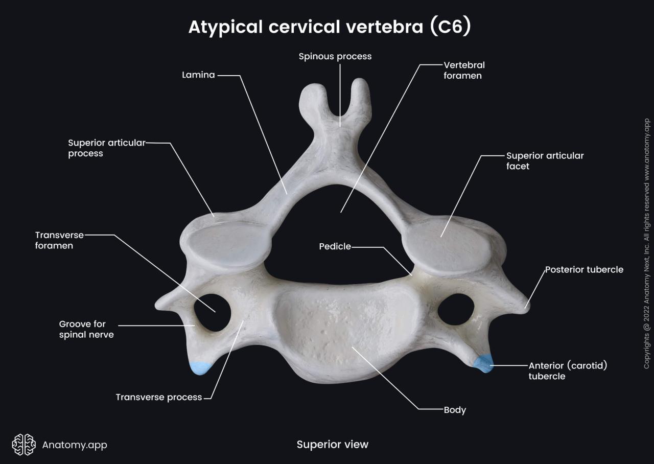
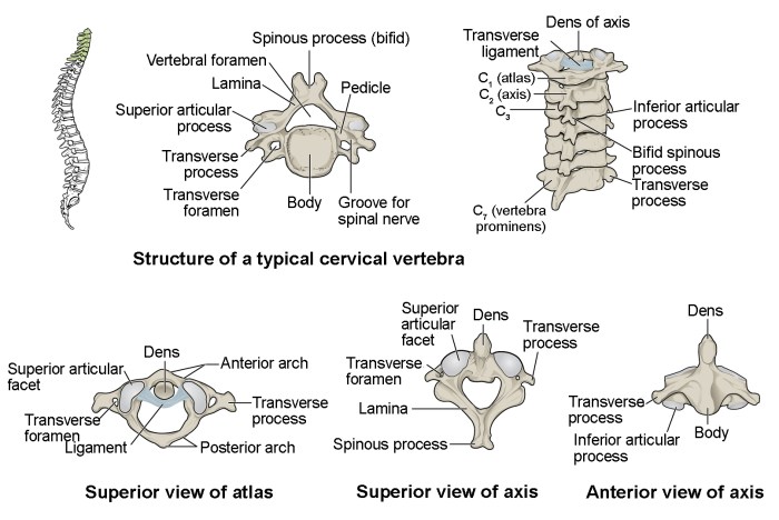
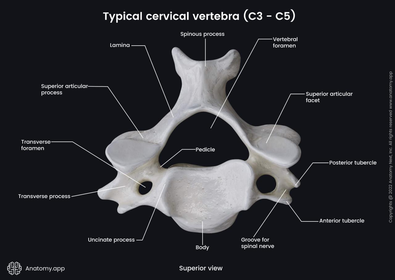
Cervical vertebrae labeled superior view: Delving into the intricate anatomy of the neck’s foundational structures, this exploration unveils the remarkable design and function of these essential vertebrae.
Positioned as the uppermost segment of the spinal column, the cervical vertebrae play a pivotal role in supporting the head, facilitating movement, and safeguarding delicate neural structures. Their intricate architecture, comprising distinct anatomical landmarks, foramina, and canals, demands a thorough understanding to appreciate their significance in human anatomy and movement.
Introduction

The cervical vertebrae, also known as the neck vertebrae, are the seven bones that make up the neck region of the spinal column. They are located between the skull and the thoracic vertebrae, and their primary purpose is to provide support, mobility, and protection for the head and neck.
Anatomy of the Cervical Vertebrae (Superior View): Cervical Vertebrae Labeled Superior View

The cervical vertebrae are relatively small and delicate compared to other vertebrae in the spinal column. They have a unique shape and structure that allows for a wide range of motion in the neck.
Vertebral Body, Cervical vertebrae labeled superior view
The vertebral body is the large, oval-shaped portion of the vertebra that forms the anterior part. It is responsible for supporting the weight of the head and neck.
Transverse Processes
The transverse processes are two bony projections that extend laterally from the vertebral body. They provide attachment points for muscles and ligaments.
Spinous Process
The spinous process is a single, midline projection that extends posteriorly from the vertebral body. It provides a lever arm for muscle attachment and helps to protect the spinal cord.
Lamina
The lamina are two thin plates of bone that extend posteriorly from the vertebral body and meet to form the roof of the vertebral foramen.
Pedicles
The pedicles are two short, thick columns of bone that connect the vertebral body to the lamina. They form the lateral walls of the vertebral foramen.
Foramina and Canals of the Cervical Vertebrae

The vertebral foramen is the large, central opening within the vertebra. It allows for the passage of the spinal cord.
Intervertebral Foramen
The intervertebral foramen is a space between two adjacent vertebrae, formed by the notches on the inferior and superior surfaces of the vertebral bodies and the pedicles. It allows for the passage of spinal nerves.
Transverse Foramen
The transverse foramen is a small opening within the transverse process. It allows for the passage of the vertebral artery and vein.
Key Questions Answered
What is the function of the transverse foramen?
The transverse foramen allows the passage of the vertebral artery and vein, as well as the sympathetic nerve fibers.
What is the clinical significance of cervical vertebrae alignment?
Proper cervical alignment is crucial for maintaining optimal spinal function, preventing nerve compression, and ensuring proper blood flow to the brain.
